A frenectomy is a simple surgical procedure that involves the removing or repositioning a band of tissue that is negatively affecting the gum tissue and/or tongue.
Two main types of frenums exist:
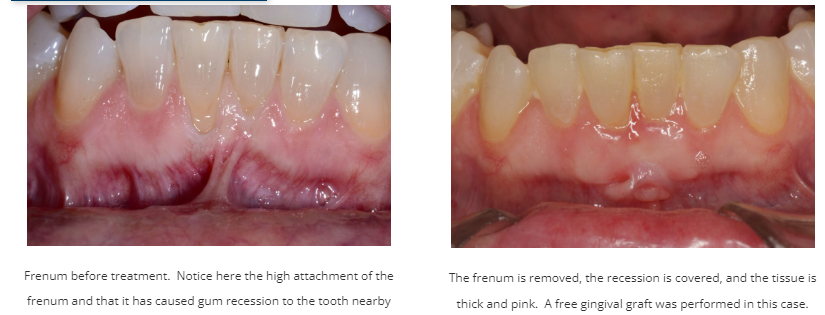
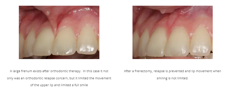
- Buccal Frenum: A band of tissue has a “high attachment” to the gum tissue. This can pull on the gums when you function, making the gum tissue prone to recession, or can make orthodontic tooth movement prone to relapse.
- Lingual Frenum: A band of tissue under the tongue can prevent proper tongue position and/or movement.
When Is A Frenectomy Needed?
Buccal Frenum
1. When the attachment of the frenum makes the gum tissue unstable. This makes the gum tissue around the frenum more mobile when you function and can make the gum tissue more prone to recession. You may notice that the gum tissue is more “white” when you stretch your lips. In the case of gum recession also being present, sometimes a gum graft removes the frenum and also helps treat the recession.
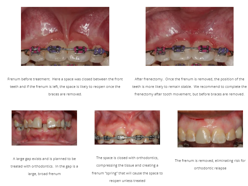
2. When a frenum makes orthodontic tooth movement unstable. When a gap in the front teeth is closed after braces, your orthodontist may recommend removal of a frenum to ensure the space remains closed.
Lingual Frenum
1. When the tongue position and/or movement is altered. The lingual frenum is a small fold of tissue that attaches the tongue to the floor of the mouth. Ideally, it should attach under the tongue at about the tongue’s midpoint, but occasionally the attachment can be unusually thick, short or tight. This is commonly known as being “tongue tied”. In infants, this condition is almost always addressed and treated. For adults, it’s common to leave untreated unless it interferes with the normal function of the tongue. It is crucial that the attachment allows the tongue to move in a way to facilitate proper speech and development of the jaws. Research suggests that if the tongue doesn’t properly move to the roof of the mouth, it can have negative effects on the development of the upper jaw and your child’s ability to breathe properly.
PERFORMING THE PROCEDURE
If it is discovered that you or child are suffering the undesirable side effects of an aberrant frenum, a frenectomy may be recommended as a permanent solution. Dr. Schaberg or Dr. Campbell will assess your needs to determine whether or not the procedure is necessary. If a frenectomy is deemed necessary it is performed in our office, typically lasting only 15 to 20 minutes. Recovery is considered complete within a matter of two weeks, during which time only mild discomfort is typically experienced. Proper compliance with post-operative instructions is important, especially when a lingual frenectomy is performed. Sometimes exercises from an orofacial myofunctional therapist or speech pathologist may be necessary in conjunction with our post-operative recommendations.